Vascularized 3D Tissue & Organ Models
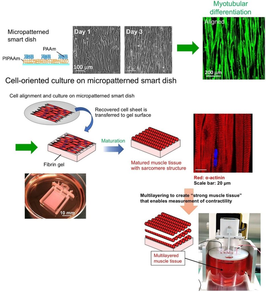
Simply repeating the layering of cell sheets will result in limited tissue thickness due to lack of oxygen and nutrients inside the tissue and accumulation of waste products. Our research group has devised a technology to create a network of blood vessels in tissues by in vivo grafting cell sheets, and finally successfully constructed thick three-dimensional tissues. To realize this technology in vitro, we are currently developing a vascular network delivery system using vascular beds and tissue perfusion bioreactors.
Lymphatic and Vascular Engineered Tissue for Lymphedema Treatment
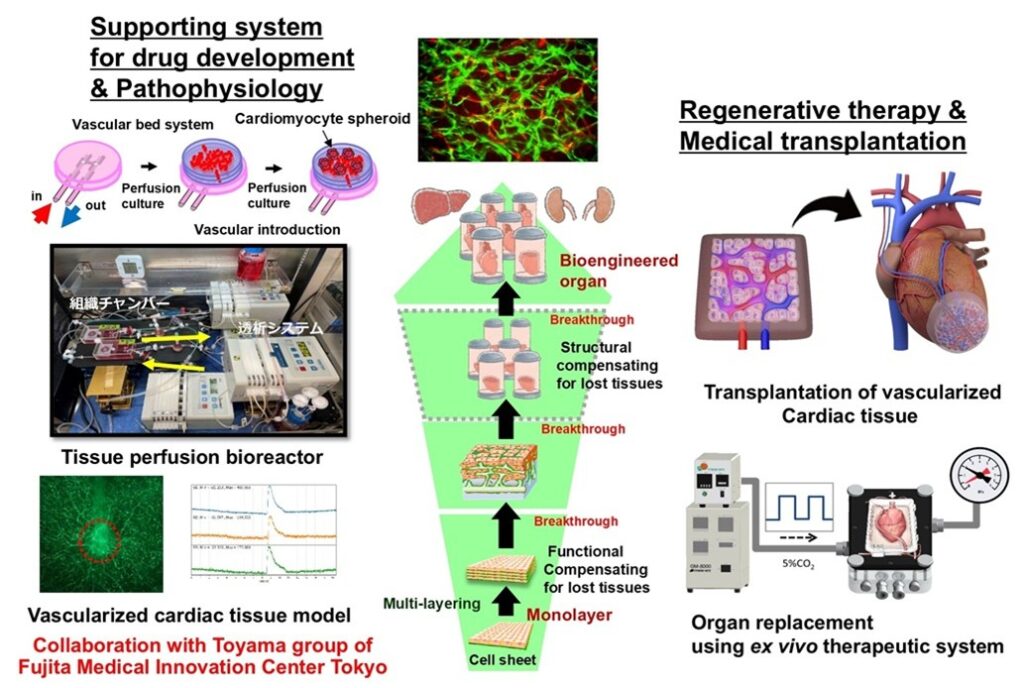
Lymphedema is a disease characterized by chronic swelling of the extremities due to excessive fluid accumulation, fibrosis of the tissues, accumulation of subcutaneous fat, decreased immune function, impaired wound healing, and increased risk of infection. In addition to changes in appearance, it may cause disability and be life-threatening. Once lymphedema develops, it often requires lifelong management, and no curative treatment has been established. Our research group works on the development of fundamental treatment for intractable lymphedema using a transplantable vascularized lymphatic tissue. The vascularized lymphatic tissue is expected to construct utilizing a perfusion culture system of co-cultured cell sheets with endothelial cell networks that construct lymphatic vessels and blood vessels, respectively. We aim to rapidly increase lymphatic capillary density by creating the transplantable lymphatic vascularized tissue that has the ability to pump lymphatic fluid, which is important for maintaining fluid homeostasis, and to drain it into veins.
Human Cardiac Tissue Model and Contractility Measurement System
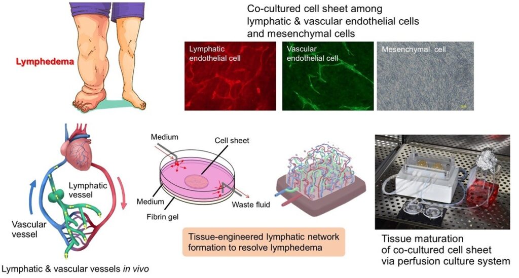
Animal models have been used in disease and drug discovery research. However, it is desired to develop tissue models that more closely resemble human organisms. Our research group has developed a system for measuring the contractile force of beating cardiac tissues generated from human iPS cell-derived cardiac cells. This system is used for evaluating drug efficacy and cardiotoxicity on commercial service in collaboration with a joint research company. We also work on the application of this system to the evaluation of contractile function of cardiac tissues for transplantation in vivo and pathological research as a model of pathological tissue.
Soft Nanomesh Sensor for Measuring Surface Potential of Self-beating Cardiac Tissues
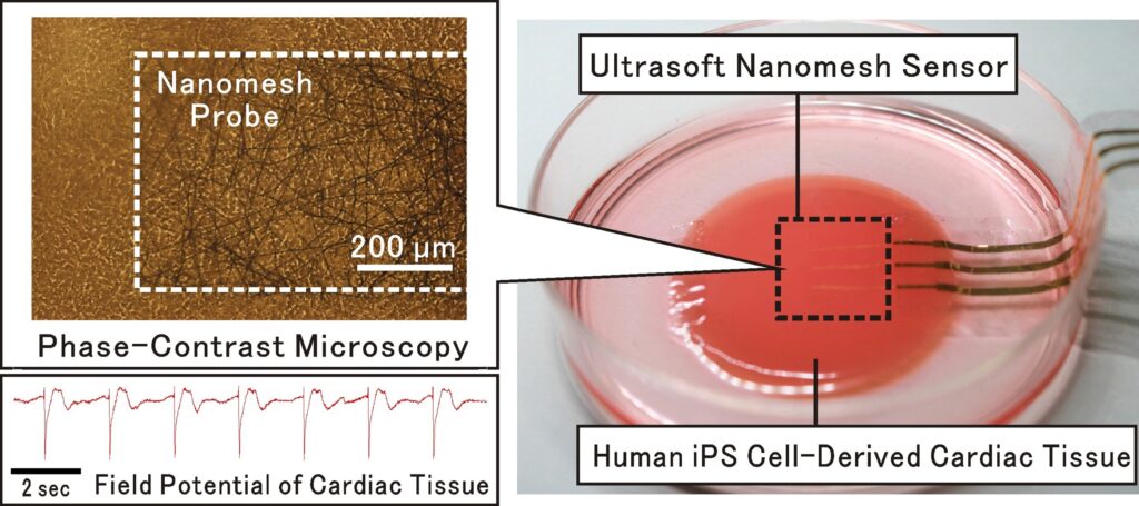
By utilizing of iPS cells and cell sheet engineering, it has become possible to create human myocardial tissue that beats as strongly like a living heart tissue. Our research group has developed the world’s first ultra-flexible nanomesh sensor that can measure the surface potential of beating human myocardial tissue, collaborated with Prof. Someya’s groups at the University of Tokyo. This measurement system can be used for drug efficacy and toxicity testing of compounds without using experimental animals, and for functional evaluation of cardiac muscle tissue to be produced for regenerative medicine. Based on this technology, research is also expected to expand into the development of highly functional implanted tissues and bio-actuators that integrate electronics.
Dome-shaped cultured myocardial tissue: Miniature heart
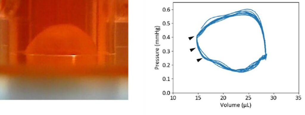
Our research group has created a dome-shaped cultured myocardial tissue (miniature heart) by utilizing cell sheets consisting of human iPS cell-derived cardiomyocytes. This miniature heart can function as a pump and circulate culture fluid just like the human heart. Furthermore, we have successfully obtained a pressure-volume diagram, which is used in clinical practice as a precise examination of cardiac function.
Human Cardiac Tissue Model with Controlled Cell Orientation
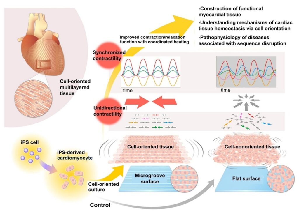
The construction of biomimicking cardiac tissues is an important issue not only for regenerative medicine, but also for applications in disease and drug discovery research and in understanding the living heart. Although the heart has a tissue structure with oriented cardiomyocytes, the effect and mechanism on the contractile and relaxing functions of the tissue as a whole have not been clarified.
Our research group has successfully produced human cardiac tissue models with controlled cardiomyocyte orientation. The oriented cardiomyocytes show unidirectional contractility-relaxation, and thus this function of the whole tissue is enhanced by promoting synchronous contraction. This finding is expected not only to be applied to the construction of various medical cardiac tissues, but also to clarify the relationship between the disorder of cell arrangement and abnormalities in contraction and relaxation property and proarrhythmic effects in the pathogenesis of cardiac diseases.
3D Human Skeletal Muscle Tissue Model and Contractility Measurement System
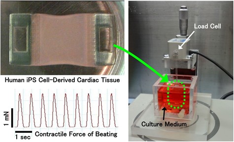
Based on cell sheet engineering, our group has successfully produced a three-dimensional human skeletal muscle tissue model that structurally and functionally mimics living muscle tissue via oriented myofiber formation. In addition, a system to measure the muscle contraction via electrical stimulation of the tissue has been also constructed. This system facilitates the real-time detection of drug effects on muscle tissue based on the contraction force change and can be applied to the development of treatments for intractable muscle diseases.
On the other hand, the living skeletal muscle receives nerve-derived signals to move, and thus any abnormality in the nerve tissue will also affect or cause abnormalities in the muscle tissue. Our group has previously shown that the neurons elongate dendrites along the same direction as the muscle fibers in the co-culture between the oriented muscle fibers and human iPS cell-derived neurons. The construction of neuro-muscular tissue with physiologically connected neuro-muscular junctions is expected to contribute to the investigation of the causes of neuro-muscular diseases and the development of treatments.